Abstract
This study aimed to evaluate the pressure exerted on mandibular soft tissues by the removable components of two different extracoronal precision attachment systems in distal extension cases (Kennedy Class II). A total of 10 patients presenting with bilateral mandibular edentulous areas were included. The first and second premolars were prepared as abutment teeth. A rigid attachment system (MK1) was applied on one side, while a resilient attachment system (ASC 52) was placed on the opposite side. Pressure measurements were obtained immediately after prosthesis placement and after six months of function. Small perforations were created in the removable components at both proximal and distal points. These areas were analyzed using an optical microscope connected to a digital imaging system, allowing measurements in microns. The findings indicated no statistically significant differences between proximal and distal points in both attachment systems immediately after insertion. However, after six months, significant differences were observed in the rigid attachment system (MK1), particularly at distal points. Within the limitations of this study, resilient attachment systems demonstrated a more favorable pressure distribution on soft tissues compared to rigid attachment system.
Keywords
Extracoronal Attachments, Kennedy Class II, Removable Part Pressure, Optical Microscope, Soft Tissue Pressure,
Precision Attachment
1. Introduction
The management of partially edentulous patients, particularly in distal extension cases, remains a complex challenge in prosthodontics. In such cases, fixed prosthetic solutions are often not feasible due to anatomical limitations or patient-related factors.
Although implant-supported restorations represent a reliable alternative, their application may be limited by insufficient bone volume, systemic conditions, or financial considerations. Therefore, removable partial dentures (RPDs) continue to play an essential role in prosthetic rehabilitation
| [1] | Burns DR, Ward JE. A review of attachments for removable partial denture design: Part 1. Classification and selection. Int J Prosthodont. 1990; 3: 98–102. |
| [2] | Burns DR, Ward JE. A review of attachments for removable partial denture design: Part 2. Treatment planning and attachment selection. Int J Prosthodont. 1990; 3: 169–174. |
[1, 2]
.
Various treatment approaches have been described for restoring missing teeth, including conventional removable dentures, cast partial dentures, attachment-retained prostheses, telescopic systems, and implant-supported options. Among these, precision attachment-retained prostheses provide a balance between function and esthetics
| [3] | Reddy RK, Thumati P, Reddy GVM. Prosthetic rehabilitation of a partially edentulous condition using extracoronal semiprecision attachment and cast partial denture: A clinical report. J Int Dent Med Res. 2013; 6(3): 113–116. |
[3]
.
Precision attachments are mechanical connectors consisting of two components: one attached to the abutment tooth or implant, and the other integrated into the prosthesis. These systems improve retention and allow controlled movement, contributing to a more favorable distribution of occlusal forces
| [4] | Nigam A, Singh A, Shekhar A, Gupta H. Precision attachments: An overview. J Dentofacial Sci. 2013; 2(4): 41–44. |
[4]
.
At the end of the 19th century, pioneers such as Parr, Peeso, and Chayes developed early attachment systems designed to combine the advantages of fixed and removable prostheses
| [5] | Preiskel HW. Precision Attachments in Prosthodontics. London: Quintessence Publishing; 1995. |
[5]
.
Extracoronal attachments, later introduced in the early 20th century, allow vertical and rotational movement, thereby reducing stress concentration on abutment teeth by transferring part of the load to the supporting tissues.
| [6] | Preiskel HW. Precision Attachments in Dentistry. 3rd ed. St. Louis: C. V. Mosby; 1979. |
| [7] | Weaver SM. Precision attachments and their advantages in relation to supporting tissues. J Am Dent Assoc. 1938; 25: 1250–1259. |
[6, 7]
.
The few retrospective studies available show a survival rate of 83.3% for 5 years, of 67.3% up to 15 years and of 50% when extrapolated to 20 years
| [8] | Kodgi A, Akulwar R, Khandare A. Precision attachment: A precise solution for distal extension cases. Glob J Res Anal. 2014; 3(7): 239–241. |
| [9] | Makkar S, Chhabra A, Khare A. Attachment-retained removable partial denture: A case report. J Clin Dent Sci. 2011; 2(2): 39–43. |
[8, 9]
.
Attachments can be applied in crown and bridge restorations, removable partial denture restorations, overdenture restorations, and implant restorations. Their clinical application and treatment planning principles have been extensively described in the literature
| [12] | Preiskel HW. Precision Attachments in Prosthodontics: Overdentures and Telescopic Prostheses. Chicago: Quintessence Publishing; 1985. |
| [13] | Feinberg E. Diagnosing and prescribing therapeutic attachment-retained partial dentures. N Y State Dent J. 1982; 48(1): 27–29. |
[12, 13]
. Free-end saddle prostheses particularly benefit from precision attachment systems because of their favorable biomechanical characteristics
| [14] | Preiskel H. Precision attachments for free-end saddle prostheses. Br Dent J. 1969; 127: 462–468. |
[14]
.
The selection of an appropriate attachment system depends on several factors, including available space, functional requirements, cost, and the biomechanical behavior of the prosthesis. Attachments can generally be classified as rigid or resilient, depending on their capacity to absorb functional stresses
| [10] | Peter ES, Darwin B. Attachments and Implants. Coesfeld, Germany: IS-Dental; 2002. |
[10]
.
Understanding the influence of different attachment systems on stress distribution in soft tissues is essential for improving clinical outcomes
| [9] | Makkar S, Chhabra A, Khare A. Attachment-retained removable partial denture: A case report. J Clin Dent Sci. 2011; 2(2): 39–43. |
| [10] | Peter ES, Darwin B. Attachments and Implants. Coesfeld, Germany: IS-Dental; 2002. |
[9, 10]
.
Recent studies have increasingly focused on the biomechanical performance of extracoronal attachments in distal extension cases
| [20] | Abd Allah DAE, Nawar NH, Abdelfattah AM. Effect of two esthetic digitally produced materials used in fabrication of extracoronal attachments on stresses induced in removable partial dentures. BMC Oral Health. 2024; 24: 760.
https://doi.org/10.1186/s12903-024-04477-2 |
| [21] | Tushar S, Singh A, Rani P, Prakash J, Sunila BS, Shivakumar GC. Prosthodontic rehabilitation with Kennedy Class I and II using extended precision attachment: Report of two cases. Cureus. 2023; 15(8): e43723.
https://doi.org/10.7759/cureus.43723 |
| [22] | Elkerdawy MW, Aboutar SNA, Fayyad AEE. Kennedy Class II problems and treatment options: A literature review. Adv Dent J. 2023; 5(3): 681–689.
https://doi.org/10.21608/adjc.2023.228891.1382 |
| [23] | Helmy MA, Kamel S, Kothayer M. Effect of different extracoronal attachment designs on biological considerations and bacterial growth in mandibular Kennedy Class II cases: A randomized clinical trial. Egypt Dent J. 2024.
https://doi.org/10.21608/edj.2024.261195.2871 |
[20-23]
.
2. Objective and Significance of the Study
This study aimed to evaluate the pressure exerted by the removable components of extracoronal precision attachments on mandibular soft tissues and to compare the pressure distribution between proximal and distal regions in Kennedy Class II cases.
Additionally, the study sought to determine which attachment type provides a more favorable biomechanical response in situations where fixed or implant-supported treatments are not feasible.
3. Materials and Methods
3.1. Study Design and Sample
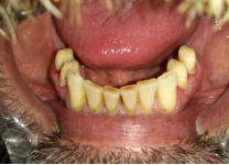
This clinical study was conducted on a total of 10 patients presenting with mandibular Kennedy Class II edentulous areas. All patients met the inclusion criteria and provided informed consent prior to participation.
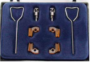
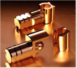
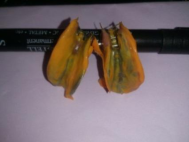
Each patient received two different extracoronal precision attachment systems: a rigid attachment system (MK1) (
Figure 1) on one side and a resilient attachment system (ASC 52) (
Figure 2) on the contralateral side. The first and second premolars were selected as abutment teeth in all cases.
Figure 1. MK1 rigid extracoronal attachment used in the study.
Figure 2. ASC 52 extracoronal attachment used in the study.
3.2. Inclusion Criteria
The inclusion criteria for patient selection were as follows:
1) Presence of unilateral distal extension edentulous area in the mandible (Kennedy Class II)
2) Availability of first and second premolars as abutment teeth on both sides
3) Adequate periodontal support (at least half of the root length embedded in bone)
4) Opposing dentition consisting of natural teeth or fixed prostheses
5) Sufficient interarch space for prosthetic rehabilitation Selection Conditions.
3.3. Materials
The materials and equipment used in this study included a turbine handpiece, diamond burs (Komet, Germany), silicone impression materials (Zhermack, Italy), glass ionomer cement (Ivoclar Vivadent), dental stone, nickel-chromium alloy, ceramic materials (IPS Classic, Ivoclar Vivadent), self-curing acrylic resin, and precision attachment systems (ASC 52 and MK1).
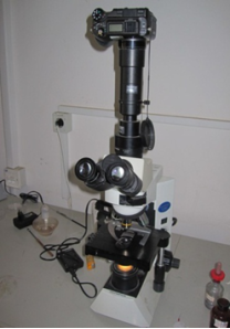
An optical microscope (Olympus CX41RF, Japan) (
Figure 3) with a maximum magnification of 500× was used, connected to a digital imaging system for measurement analysis. A calibrated micrometric ruler (
Figure 4) was utilized to convert pixel-based measurements into microns.
Figure 3. Optical microscope system used for image acquisition and measurement.
Figure 4. Calibration ruler used for microscopic measurements.
3.4. Prosthetic Procedure
The prosthetic procedure was carried out in several stages. The abutment teeth (first and second premolars) were prepared to receive metal-ceramic crowns. Following tooth preparation, impressions were taken and casts were fabricated (
Figure 5).
Figure 5. Preparation of abutment teeth for metal-ceramic crowns.
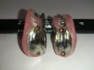
The female components of the attachments were incorporated into the crowns during the waxing stage and subsequently cast using a nickel-chromium alloy. After ceramic application and finishing, the crowns were clinically evaluated (
Figure 6).
Figure 6. Ceramic crowns and attachment integration during crown fabrication.
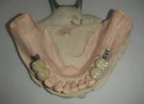
A new impression was then taken with the crowns in place to obtain a working cast. The removable prosthetic components were fabricated using self-curing acrylic resin and metal framework, followed by the incorporation of artificial teeth and gingival structures (
Figure 7).
Figure 7. Fabrication of the removable prosthetic component.
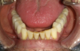
Finally, the prostheses were inserted intraorally, ensuring proper engagement between male and female components. Cementation was performed using glass ionomer cement (
Figure 8).
Figure 8. Clinical placement of prosthesis with attachment system.
3.5. Pressure Measurement Procedure
Pressure evaluation was conducted at two time intervals:
1) Immediately after prosthesis insertion
2) After six months of clinical use
Standardized perforations (1 mm depth) were created at the proximal and distal regions of the removable components of the attachments. (
Figure 9).
Figure 9. Standardized perforation created at proximal and distal points for pressure measurement.
Figure 10. Silicone impression taken under functional loading conditions.
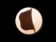
Under functional loading conditions, silicone impressions were taken while patients were instructed to bite firmly. The resulting samples were sectioned horizontally using two parallel blades spaced 0.5 mm apart to obtain uniform microscopic sections (
Figure 10).
The sections were analyzed under 100× magnification using the optical microscope. Digital images were captured and processed using specialized software (
Figure 11).
Figure 11. Microscopic section of perforation area.
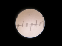
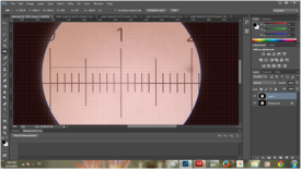
A calibration process was performed using a micrometric ruler to establish the relationship between pixel measurements and real distances. Horizontal distances within the samples were measured at three standardized points and converted into microns (
Figure 12)
3.6. Statistical Analysis
All data were recorded and analyzed using SPSS software (Version 22.0). Mean values were calculated for all measurements.
Figure 12. Calibration process for converting pixel measurements to microns.
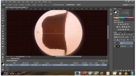
Figure 13. Digital measurement of pressure-related space using software analysis.
The Wilcoxon signed-rank test was used to compare related samples (
Figure 13). Statistical significance was set at p < 0.05.
4. Results
Statistical analysis was performed using the Wilcoxon signed-rank test to evaluate differences between proximal and distal measurement points within each attachment system, as well as differences between the two attachment types at different time intervals.
4.1. Comparison Between Proximal and Distal Points Immediately After Insertion
No statistically significant difference was observed between proximal and distal measurement points for either attachment system (ASC 52 and MK1) immediately after prosthesis insertion (p > 0.05).
These findings indicate that both attachment systems demonstrated a similar pressure distribution pattern at the time of initial placement.
4.2. Comparison Between Proximal and Distal Points After Six Months
After six months of clinical use, no statistically significant difference was found between proximal and distal measurement points in the resilient attachment system (ASC 52) (p > 0.05).
In contrast, a statistically significant difference was observed in the rigid attachment system (MK1) between proximal and distal points (p < 0.05). The distal region exhibited lower measured space values, indicating increased pressure on the soft tissues.
4.3. Comparison Between Attachment Systems
No statistically significant difference was found between the two attachment systems in distal measurements immediately after prosthesis insertion (p > 0.05).
However, after six months, a statistically significant difference was observed between the distal measurements of the two systems (p < 0.05). The rigid attachment system (MK1) demonstrated higher pressure on the distal extension area compared to the resilient attachment system (ASC 52).
Overall, the results indicate that while both attachment systems perform similarly at the time of insertion, the resilient attachment system maintains a more favorable pressure distribution over time, particularly in distal regions.
5. Discussion
Distal extension cases (Kennedy Class II) present significant biomechanical challenges due to the difference in support between natural teeth and soft tissues. This discrepancy often results in uneven stress distribution during functional loading, potentially compromising the longevity of both the prosthesis and supporting structures
| [11] | Ashish R, Jacob M, Padma A. Attachment-retained distal extension cast partial denture: A case report. Int J Prosthodont Restor Dent. 2012; 2(3): 101–107. |
| [17] | Sahin V, Akaltan F, Parnas L. Effects of retainer type and rigidity on stress distribution in telescopic-retained removable partial dentures. J Dent Sci. 2012; 7: 7–13. |
[11, 17]
.
The results of the present study demonstrated that both rigid and resilient attachment systems exhibited similar pressure distribution patterns immediately after prosthesis insertion. This finding may be attributed to the initial adaptation phase, during which functional loading has not yet significantly influenced the biomechanical behavior of the prosthesis.
However, after six months of clinical use, a statistically significant difference was observed in the rigid attachment system (MK1), particularly at distal regions. The reduced space values in these areas indicate increased pressure on the underlying soft tissues. This suggests that rigid attachments tend to transmit occlusal forces more directly to the distal extension base, leading to stress concentration over time
| [18] | Wang H, Zhang Y, Yao D, Chen J. Effects of rigid and nonrigid extracoronal attachments on supporting tissues in extension base removable partial dentures: A nonlinear finite element study. J Prosthet Dent. 2011; 105(5): 338–346. |
| [19] | Nishimura RD, Ochiai KT, Caputo AA, Jeong CM. Photoelastic stress analysis of load transfer to implants and natural teeth comparing rigid and semirigid connectors. J Prosthet Dent. 1999; 81: 696–703. |
[18, 19]
.
In contrast, the resilient attachment system (ASC 52) maintained a more uniform pressure distribution, with no statistically significant differences between proximal and distal regions after six months. This behavior can be explained by the design of resilient attachments, which allows controlled vertical and rotational movement, thereby acting as a stress-breaking mechanism
| [17] | Sahin V, Akaltan F, Parnas L. Effects of retainer type and rigidity on stress distribution in telescopic-retained removable partial dentures. J Dent Sci. 2012; 7: 7–13. |
| [18] | Wang H, Zhang Y, Yao D, Chen J. Effects of rigid and nonrigid extracoronal attachments on supporting tissues in extension base removable partial dentures: A nonlinear finite element study. J Prosthet Dent. 2011; 105(5): 338–346. |
[17, 18]
.
These findings are consistent with previous studies. Wang et al. reported that non-rigid extracoronal attachments significantly reduce stress concentration on supporting tissues, as demonstrated through finite element analysis
| [18] | Wang H, Zhang Y, Yao D, Chen J. Effects of rigid and nonrigid extracoronal attachments on supporting tissues in extension base removable partial dentures: A nonlinear finite element study. J Prosthet Dent. 2011; 105(5): 338–346. |
[18]
. Similarly, Nishimura et al. showed that semi-rigid connectors improve load transfer and reduce stress on abutment teeth and surrounding tissues compared to rigid systems
| [19] | Nishimura RD, Ochiai KT, Caputo AA, Jeong CM. Photoelastic stress analysis of load transfer to implants and natural teeth comparing rigid and semirigid connectors. J Prosthet Dent. 1999; 81: 696–703. |
[19]
.
From a clinical perspective, excessive pressure in distal extension areas may lead to mucosal irritation, residual ridge resorption, and patient discomfort over time
| [15] | Picton DC, Willis DJ. Viscoelastic properties of the periodontal membrane and mucous membrane. J Prosthet Dent. 1978; 40: 263–272. |
| [16] | Kapur KK, Deupree R, Dent RJ, Hasse AL. A randomized clinical trial of two basic removable partial denture designs. Part I: Comparison of five-year success rates and periodontal health. J Prosthet Dent. 1994; 72(3): 268–282. |
[15, 16]
. Therefore, the ability of resilient attachment systems to distribute functional loads more evenly represents a clear clinical advantage.
The findings of this study support the growing body of evidence suggesting that attachment flexibility plays a crucial role in stress modulation and long-term prosthetic success
| [17] | Sahin V, Akaltan F, Parnas L. Effects of retainer type and rigidity on stress distribution in telescopic-retained removable partial dentures. J Dent Sci. 2012; 7: 7–13. |
| [18] | Wang H, Zhang Y, Yao D, Chen J. Effects of rigid and nonrigid extracoronal attachments on supporting tissues in extension base removable partial dentures: A nonlinear finite element study. J Prosthet Dent. 2011; 105(5): 338–346. |
| [19] | Nishimura RD, Ochiai KT, Caputo AA, Jeong CM. Photoelastic stress analysis of load transfer to implants and natural teeth comparing rigid and semirigid connectors. J Prosthet Dent. 1999; 81: 696–703. |
| [23] | Helmy MA, Kamel S, Kothayer M. Effect of different extracoronal attachment designs on biological considerations and bacterial growth in mandibular Kennedy Class II cases: A randomized clinical trial. Egypt Dent J. 2024.
https://doi.org/10.21608/edj.2024.261195.2871 |
[17-19, 23]
.
However, this study has certain limitations, including the relatively small sample size and the limited follow-up period. Future studies with larger populations and longer observation periods are recommended to further validate these results and enhance their generalizability.
Despite these limitations, the present study provides valuable clinical insight into the biomechanical behavior of extracoronal attachment systems and highlights the advantages of resilient designs in distal extension cases.
6. Conclusions
Within the limitations of this clinical study, extracoronal precision attachments can be considered a reliable treatment option for the rehabilitation of mandibular Kennedy Class II cases.
The findings indicate that rigid attachment systems tend to generate higher pressure on distal soft tissues over time, whereas resilient attachment systems provide a more favorable and balanced distribution of functional loads.
From a clinical standpoint, the use of resilient attachments may contribute to improved patient comfort, reduced risk of soft tissue damage, and enhanced long-term prosthetic performance in distal extension cases.
Therefore, when biomechanical considerations are prioritized, resilient extracoronal attachment systems should be preferred over rigid designs in Kennedy Class II rehabilitations.
These findings may help clinicians select the most appropriate attachment system for distal extension cases.
Abbreviations
RPD | Removable Partial Denture |
ASC 52 | Resilient Extracoronal Attachment System |
MK1 | Rigid Extracoronal Attachment System |
SPSS | Statistical Package for the Social Sciences |
mm | Millimeter |
µm | Micrometer |
Author Contributions
Deniz Suleiman: Conceptualization, Methodology, Investigation, Data Curation, Formal Analysis, Visualization, Writing – original draft, Writing – review & editing
Conflicts of Interest
The author declares no conflicts of interest.
References
| [1] |
Burns DR, Ward JE. A review of attachments for removable partial denture design: Part 1. Classification and selection. Int J Prosthodont. 1990; 3: 98–102.
|
| [2] |
Burns DR, Ward JE. A review of attachments for removable partial denture design: Part 2. Treatment planning and attachment selection. Int J Prosthodont. 1990; 3: 169–174.
|
| [3] |
Reddy RK, Thumati P, Reddy GVM. Prosthetic rehabilitation of a partially edentulous condition using extracoronal semiprecision attachment and cast partial denture: A clinical report. J Int Dent Med Res. 2013; 6(3): 113–116.
|
| [4] |
Nigam A, Singh A, Shekhar A, Gupta H. Precision attachments: An overview. J Dentofacial Sci. 2013; 2(4): 41–44.
|
| [5] |
Preiskel HW. Precision Attachments in Prosthodontics. London: Quintessence Publishing; 1995.
|
| [6] |
Preiskel HW. Precision Attachments in Dentistry. 3rd ed. St. Louis: C. V. Mosby; 1979.
|
| [7] |
Weaver SM. Precision attachments and their advantages in relation to supporting tissues. J Am Dent Assoc. 1938; 25: 1250–1259.
|
| [8] |
Kodgi A, Akulwar R, Khandare A. Precision attachment: A precise solution for distal extension cases. Glob J Res Anal. 2014; 3(7): 239–241.
|
| [9] |
Makkar S, Chhabra A, Khare A. Attachment-retained removable partial denture: A case report. J Clin Dent Sci. 2011; 2(2): 39–43.
|
| [10] |
Peter ES, Darwin B. Attachments and Implants. Coesfeld, Germany: IS-Dental; 2002.
|
| [11] |
Ashish R, Jacob M, Padma A. Attachment-retained distal extension cast partial denture: A case report. Int J Prosthodont Restor Dent. 2012; 2(3): 101–107.
|
| [12] |
Preiskel HW. Precision Attachments in Prosthodontics: Overdentures and Telescopic Prostheses. Chicago: Quintessence Publishing; 1985.
|
| [13] |
Feinberg E. Diagnosing and prescribing therapeutic attachment-retained partial dentures. N Y State Dent J. 1982; 48(1): 27–29.
|
| [14] |
Preiskel H. Precision attachments for free-end saddle prostheses. Br Dent J. 1969; 127: 462–468.
|
| [15] |
Picton DC, Willis DJ. Viscoelastic properties of the periodontal membrane and mucous membrane. J Prosthet Dent. 1978; 40: 263–272.
|
| [16] |
Kapur KK, Deupree R, Dent RJ, Hasse AL. A randomized clinical trial of two basic removable partial denture designs. Part I: Comparison of five-year success rates and periodontal health. J Prosthet Dent. 1994; 72(3): 268–282.
|
| [17] |
Sahin V, Akaltan F, Parnas L. Effects of retainer type and rigidity on stress distribution in telescopic-retained removable partial dentures. J Dent Sci. 2012; 7: 7–13.
|
| [18] |
Wang H, Zhang Y, Yao D, Chen J. Effects of rigid and nonrigid extracoronal attachments on supporting tissues in extension base removable partial dentures: A nonlinear finite element study. J Prosthet Dent. 2011; 105(5): 338–346.
|
| [19] |
Nishimura RD, Ochiai KT, Caputo AA, Jeong CM. Photoelastic stress analysis of load transfer to implants and natural teeth comparing rigid and semirigid connectors. J Prosthet Dent. 1999; 81: 696–703.
|
| [20] |
Abd Allah DAE, Nawar NH, Abdelfattah AM. Effect of two esthetic digitally produced materials used in fabrication of extracoronal attachments on stresses induced in removable partial dentures. BMC Oral Health. 2024; 24: 760.
https://doi.org/10.1186/s12903-024-04477-2
|
| [21] |
Tushar S, Singh A, Rani P, Prakash J, Sunila BS, Shivakumar GC. Prosthodontic rehabilitation with Kennedy Class I and II using extended precision attachment: Report of two cases. Cureus. 2023; 15(8): e43723.
https://doi.org/10.7759/cureus.43723
|
| [22] |
Elkerdawy MW, Aboutar SNA, Fayyad AEE. Kennedy Class II problems and treatment options: A literature review. Adv Dent J. 2023; 5(3): 681–689.
https://doi.org/10.21608/adjc.2023.228891.1382
|
| [23] |
Helmy MA, Kamel S, Kothayer M. Effect of different extracoronal attachment designs on biological considerations and bacterial growth in mandibular Kennedy Class II cases: A randomized clinical trial. Egypt Dent J. 2024.
https://doi.org/10.21608/edj.2024.261195.2871
|
Cite This Article
-
APA Style
Suleiman, D. (2026). A Clinical Study to Assess the Pressure of the Removable Part of the Precision Attachments on the Soft Tissues in Cases of Kennedy Class II. International Journal of Biomedical Engineering and Clinical Science, 12(1), 7-13. https://doi.org/10.11648/j.ijbecs.20261201.12
 Copy
|
Copy
|
 Download
ACS Style
Suleiman, D. A Clinical Study to Assess the Pressure of the Removable Part of the Precision Attachments on the Soft Tissues in Cases of Kennedy Class II. Int. J. Biomed. Eng. Clin. Sci. 2026, 12(1), 7-13. doi: 10.11648/j.ijbecs.20261201.12
Copy
|
Download
AMA Style
Suleiman D. A Clinical Study to Assess the Pressure of the Removable Part of the Precision Attachments on the Soft Tissues in Cases of Kennedy Class II. Int J Biomed Eng Clin Sci. 2026;12(1):7-13. doi: 10.11648/j.ijbecs.20261201.12
Copy
|
Download
-
@article{10.11648/j.ijbecs.20261201.12,
  author = {Deniz Suleiman},
  title = {A Clinical Study to Assess the Pressure of the Removable Part of the Precision Attachments on the Soft Tissues in Cases of Kennedy Class II},
  journal = {International Journal of Biomedical Engineering and Clinical Science},
  volume = {12},
  number = {1},
  pages = {7-13},
  doi = {10.11648/j.ijbecs.20261201.12},
  url = {https://doi.org/10.11648/j.ijbecs.20261201.12},
  eprint = {https://article.sciencepublishinggroup.com/pdf/10.11648.j.ijbecs.20261201.12},
  abstract = {This study aimed to evaluate the pressure exerted on mandibular soft tissues by the removable components of two different extracoronal precision attachment systems in distal extension cases (Kennedy Class II). A total of 10 patients presenting with bilateral mandibular edentulous areas were included. The first and second premolars were prepared as abutment teeth. A rigid attachment system (MK1) was applied on one side, while a resilient attachment system (ASC 52) was placed on the opposite side. Pressure measurements were obtained immediately after prosthesis placement and after six months of function. Small perforations were created in the removable components at both proximal and distal points. These areas were analyzed using an optical microscope connected to a digital imaging system, allowing measurements in microns. The findings indicated no statistically significant differences between proximal and distal points in both attachment systems immediately after insertion. However, after six months, significant differences were observed in the rigid attachment system (MK1), particularly at distal points. Within the limitations of this study, resilient attachment systems demonstrated a more favorable pressure distribution on soft tissues compared to rigid attachment system.},
 year = {2026}
}
Copy
|
Download
-
TY  - JOUR
T1  - A Clinical Study to Assess the Pressure of the Removable Part of the Precision Attachments on the Soft Tissues in Cases of Kennedy Class II
AU  - Deniz Suleiman
Y1  - 2026/06/10
PY  - 2026
N1  - https://doi.org/10.11648/j.ijbecs.20261201.12
DO  - 10.11648/j.ijbecs.20261201.12
T2  - International Journal of Biomedical Engineering and Clinical Science
JF  - International Journal of Biomedical Engineering and Clinical Science
JO  - International Journal of Biomedical Engineering and Clinical Science
SP  - 7
EP  - 13
PB  - Science Publishing Group
SN  - 2472-1301
UR  - https://doi.org/10.11648/j.ijbecs.20261201.12
AB  - This study aimed to evaluate the pressure exerted on mandibular soft tissues by the removable components of two different extracoronal precision attachment systems in distal extension cases (Kennedy Class II). A total of 10 patients presenting with bilateral mandibular edentulous areas were included. The first and second premolars were prepared as abutment teeth. A rigid attachment system (MK1) was applied on one side, while a resilient attachment system (ASC 52) was placed on the opposite side. Pressure measurements were obtained immediately after prosthesis placement and after six months of function. Small perforations were created in the removable components at both proximal and distal points. These areas were analyzed using an optical microscope connected to a digital imaging system, allowing measurements in microns. The findings indicated no statistically significant differences between proximal and distal points in both attachment systems immediately after insertion. However, after six months, significant differences were observed in the rigid attachment system (MK1), particularly at distal points. Within the limitations of this study, resilient attachment systems demonstrated a more favorable pressure distribution on soft tissues compared to rigid attachment system.
VL  - 12
IS  - 1
ER  -
Copy
|
Download